Leica ARveo microscopes

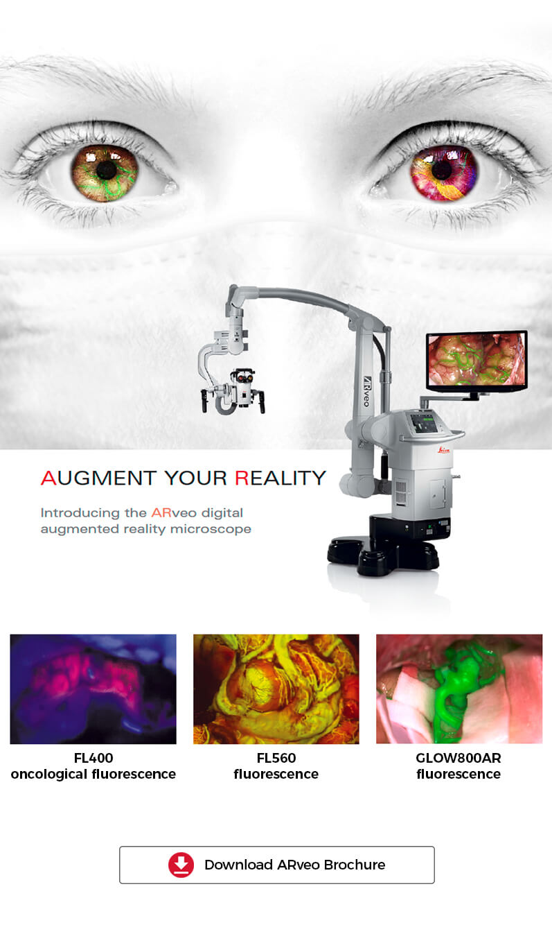
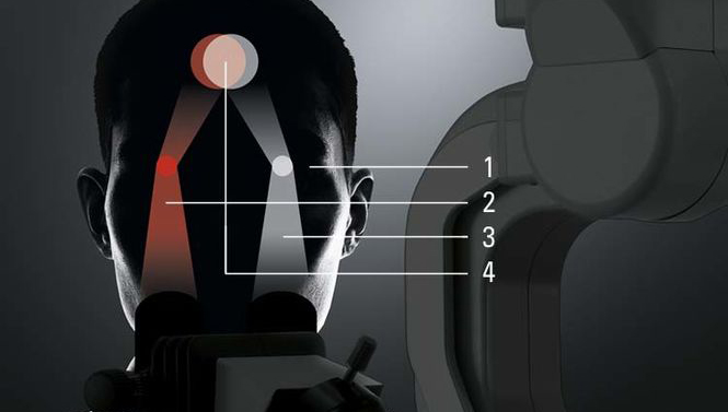
 Image injection module CaptiView
To make precise, confident decisions during critical cranial surgery, you require multiple pieces of visual data for a full picture of the surgical site.
What if you could keep your eyes and focus on your patient with full confidence that you are seeing all supporting visual information with high resolution and contrast?
With CaptiView image injection this can be your new OR experience! View FL800 fluorescence, GLOW800 Augmented Reality fluorescence, microscope information, endoscope images, and data from Image Guided Surgery (IGS) or neuronavigation systems directly in the microscope eyepieces.
Image injection module CaptiView
To make precise, confident decisions during critical cranial surgery, you require multiple pieces of visual data for a full picture of the surgical site.
What if you could keep your eyes and focus on your patient with full confidence that you are seeing all supporting visual information with high resolution and contrast?
With CaptiView image injection this can be your new OR experience! View FL800 fluorescence, GLOW800 Augmented Reality fluorescence, microscope information, endoscope images, and data from Image Guided Surgery (IGS) or neuronavigation systems directly in the microscope eyepieces.
Blue Light Fluorescence ModuleLeica FL400
FL400 fluorescence with 5-ALA allows differentiation of tumor tissue from healthy brain tissue to support precise resection.
When resecting a malignant glioma, maximum removal of tumor cells with minimal impact to brain tissue is key to an optimal patient outcome. This is where FL400 fluorescence can provide support. In combination with the active substance 5 aminolevulinic acid (5-ALA), it helps distinguish both the bulk and margins of the tumor more easily. More visual information helps you to confidently perform a resection.

Fluorescence Module Leica FL560
Visualizing certain anatomical and physiological features during neurosurgery can be challenging under white light or near infrared ICG fluorescence.
The FL560 fluorescence module enables simultaneous, real-time observation of both non-fluorescent tissue and fluorescent areas, with clear differentiation and contrast.

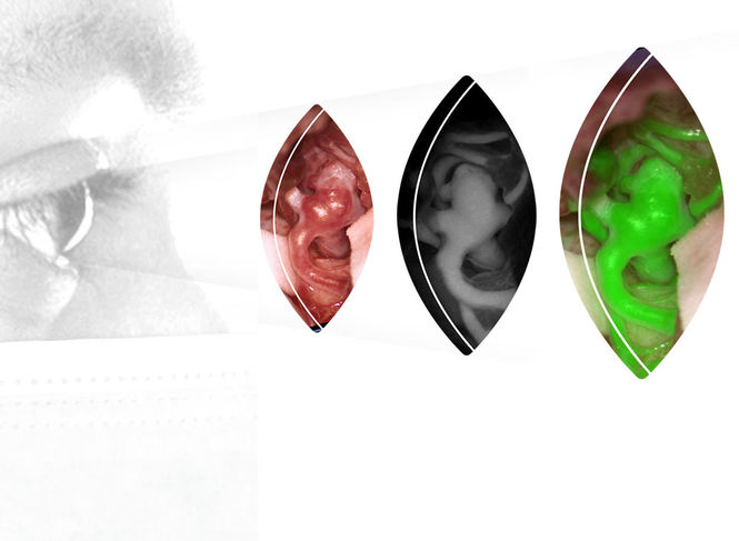
GLOW800 – Augmented Reality Fluorescence
Whether clipping an aneurysm, removing an AVM, performing a microvascular decompression or a bypass, your ability to clearly view and assess cerebral anatomy and blood flow is critical for an optimal outcome.
With GLOW800 augmented reality (AR) fluorescence and ICG you can observe cerebral anatomy in natural color, augmented by real-time vascular flow, with full depth perception.
No more interrupting surgery to switch between the natural microscope image and a flat black and white near infrared video. No more mental gymnastics to recall and reconcile the different views.
Experience the benefits of one augmented view during vascular neurosurgery for enhanced confidence in your surgical intervention.
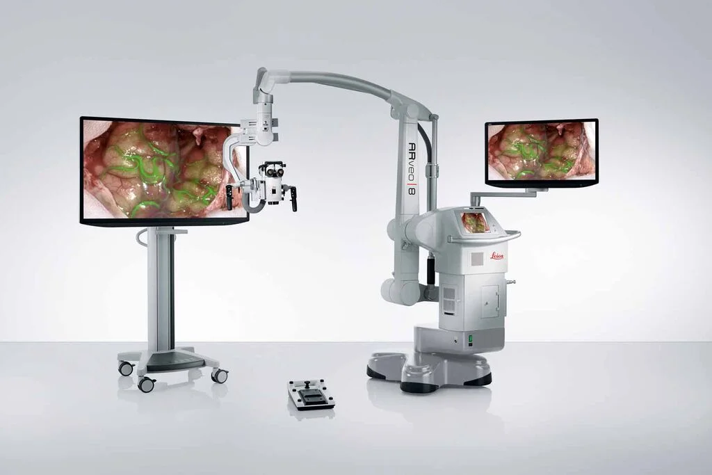
ARveo 8 Digital Visualization Microscope
ARveo 8 unites information from AR fluorescence, IGS systems, and endoscopic image feeds providing an enhanced visualization for more informed and precise neurosurgery.
With ultra-fast processing and an intuitive graphical user interface the ARveo 8 digital visualization microscope helps to enhance efficiency across the entire team.
Leica Microsystems’ EnhancePath concept for future upgradeability and system compatibility of the ARveo 8 provides a seamless way to evolve into the digital future of neurosurgery, smoothly and confidently.
Empower your decision-making with Augmented Reality
The ARveo microscope integrates the supplementary imaging information you need to make precise, confident decisions. Enhance your understanding with IGS data and our propriety GLOW Augmented Reality (AR) technology.
Building on a decade of leadership in fluorescence imaging, GLOW AR is ready to revolutionize the way you navigate your most challenging neurosurgical procedures.
A sophisticated imaging sensor and algorithms capture, optimize, and combine multiple spectral bands of visible and fluorescent light. The result is faithful, natural coloring of tissue and accurate representation of fluorescence intensity.
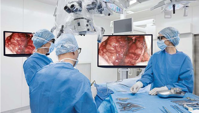
3D MONITOR VIEWING
Continue to work with the full depth perception and high resolution you require without needing to look through the eyepieces.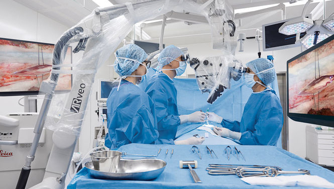
3D VIEW SHARING
Shared 3D viewing on large 4K monitors helps support workflow by enabling your whole team to follow your every delicate move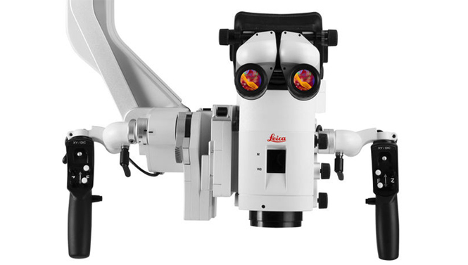
CAPTIVIEW GLOW AR DISPLAY
With CaptiView image injection you see the imaging information you need in the highest resolution, directly in the eyepieces.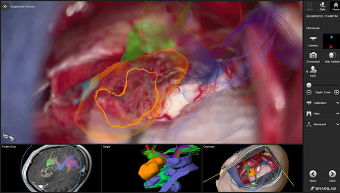
IMAGE GUIDED SURGERY
View images from IGS systems and endoscopes via eyepieces or 4K 3D screens and further augment your surgical insight. Activate the additional information display with just one touch of the microscope handle.
ENDOSCOPE IMAGING
The ARveo can display and record endoscope images from leading brands such as Aesculap